Harris View Calcaneus Fracture, Extensile Open Reduction And Internal Fixation Of Intra Articular Joint Calcaneus Fractures Musculoskeletal Key
Harris view calcaneus fracture Indeed recently is being hunted by consumers around us, maybe one of you. People now are accustomed to using the net in gadgets to see video and image data for inspiration, and according to the title of the post I will discuss about Harris View Calcaneus Fracture.
- Http Www Aamj Eg Net Journals Pdf 2159 Pdf
- Calcaneal Fractures
- Calcaneus And Talus Fractures Video Library Ota Online Trauma Access Youtube
- Calcaneal Fractures
- Foot Xray Eorif
- Simple Undisplaced Fracture Of The Calcaneus Body
Find, Read, And Discover Harris View Calcaneus Fracture, Such Us:
- Http Ota Org Media 299078 5 Calcaneus Fractures Pdf
- Plain Radiography Part I Clinical Emergency Radiology
- Mcglamry Ch 107 Calcaneal Fractures Mcglamry S Flashcards Memorang
- Calcaneus Fractures Trauma Orthobullets
- Calcaneum Fracture Pathoanatomy Various Fracture Pattern
If you are looking for Joe Biden Press you've arrived at the perfect location. We ve got 104 graphics about joe biden press including images, pictures, photos, backgrounds, and more. In such web page, we also have variety of graphics out there. Such as png, jpg, animated gifs, pic art, logo, black and white, translucent, etc.
Bilateral calcaneal fractures are seen in 5 9.

Joe biden press. A lateral and axial view of the contralateral foot should be obtained for comparison. The right image has been annotated to show bohler. Fractures of the calcaneus are common and account for approximately 60 of tarsal injuries.
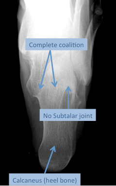
Axial harris views of both calcanei show a left calcaneal fracture. All patients with calcaneus fractures should undergo axial computed tomography ct scan of the foot. Angle between line along lateral margin of posterior facet line anterior to beak of calcaneus.
Dislocated peroneal tendons are usually treated surgically when they occur with a calcaneus fracture. Yet there remain many institutions especially in rural areas where ct is not readily available. Ap view of the foot exhibits secondary fracture lines into the anterior process and incongruity of the calcaneocuboid joint.
Standard imaging of the ankle and foot may also be indicated if you suspect associated injuries to these structures. As technology advances computed tomography ct has widely been used 1 to better visualize and characterize calcaneum fragment displacements and fracture lines. The calcaneus is the most commonly fractured tarsal bone and accounts for about 2 of all fractures.
A 55 year old male sustained a sanders iv intra articular calcaneus fracture two years ago that was treated nonoperatively. They result from an axial load and may demonstrate varying severity and location of fracture type. Calcaneal fractures can be divided broadly into two types depending on whether there is articular involvement of the subtalar joint 278.
The calcaneus axial view is part of the two view calcaneus series assessing the talocalcaneal joint and plantar aspects of the calcaneus. The calcaneus is the most commonly fractured tarsal bone and accounts for about 2 of all fractures 2 and 60 of all tarsal fractures 3. Note the fracture line divides the calcaneus into anteromedial and posterolateral fragments.
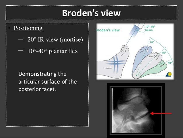
Normal lateral calcaneal xrs. The harris view is obtained with the foot in maximal dorsiflexion and the x ray beam angled 450 cephalad. Axial view harris beath visualizes tuberosity position varus valgus primary fracture line calcaneal height loss calcaneal widening and a portion of the posterior facet.
Obtain plain radiographs initially. Typically a lateral x ray demonstrating the foot from the side figure 4 as well as an axillary heel view harris view showing an end on view of the heel are taken. Measured on lateral view.
Visualizes tuberosity fragment widening shortening and varus positioning. Advances in cross sectional imaging particularly in computed tomography ct have given this modality an important role in identifying and characterizing calcaneal fractures.
More From Joe Biden Press
- Joe Biden Rally In Michigan 2020
- Joe Biden Maryland
- Kamala Harris Email Address
- Biden Vice President Pick Odds
- Doug Emhoff Ex Wife
Incoming Search Terms:
- Calcaneus Fracture In Children Springerlink Doug Emhoff Ex Wife,
- Calcaneal Fractures Doug Emhoff Ex Wife,
- Calcaneus Fractures Trauma Orthobullets Doug Emhoff Ex Wife,
- Yelena Bogdan Md Facs Faaos On Twitter I Ve Always Found Calcaneus Fractures Difficult To Image Intraoperatively This Is My Way To Get The Axial Harris View Xray From Internet Foot Guys Share Your Doug Emhoff Ex Wife,
- Calcaneal Fractures Doug Emhoff Ex Wife,
- The Magneto View A Simple Method For Obtaining Intraoperative Axial Radiographs Of The Calcaneus Doug Emhoff Ex Wife,


