Harris View Heel, The Magneto View A Simple Method For Obtaining Intraoperative Axial Radiographs Of The Calcaneus
Harris view heel Indeed lately has been sought by consumers around us, perhaps one of you. People now are accustomed to using the internet in gadgets to view image and video information for inspiration, and according to the name of this post I will discuss about Harris View Heel.
- Calcaneus Fractures Trauma Orthobullets
- Asos Design Harris Barely There Heeled Sandals Asos
- Calcaneal Fractures
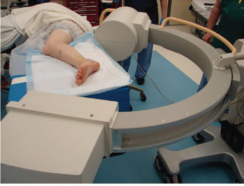
- The Magneto View A Simple Method For Obtaining Intraoperative Axial Radiographs Of The Calcaneus
- The Magneto View A Simple Method For Obtaining Intraoperative Axial Radiographs Of The Calcaneus
- Imaging Of The Foot And Ankle Musculoskeletal Key
Find, Read, And Discover Harris View Heel, Such Us:
- Xray Projections Of The Calcaneus Youtube
- Calcaneal Fractures Where Are We Now Springerlink
- Background The Bone School
- Pdf Neurovascular Structures At Risk With Curved Retrograde Ttc Fusion Nails
- Radiology In Foot And Ankle Musculoskeletal Key
If you re searching for Douglas Emhoff Ex Wife Kirsten you've come to the right place. We ve got 104 graphics about douglas emhoff ex wife kirsten adding images, photos, pictures, backgrounds, and more. In such webpage, we additionally provide number of graphics out there. Such as png, jpg, animated gifs, pic art, logo, black and white, translucent, etc.

The Magneto View A Simple Method For Obtaining Intraoperative Axial Radiographs Of The Calcaneus Douglas Emhoff Ex Wife Kirsten
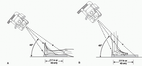
5 an axial view of the calcaneus is obtained with the x ray source posterior to the heel and tilted caudally 450 with respect to the long axis of the foot.

Douglas emhoff ex wife kirsten. Sesamoid view. Bilateral calcaneal fractures are seen in 5 9. As applied to calcaneal.
She has pain throughout the day that worsens with prolonged weight bearing. Otherwise it may need surgery to realign the bone. If the fracture is not misaligned and no gap in the bone it can be treated with a cast.
Patient standing on x ray plate with knees in slight flexion central ray angles varies from 350 to 450 aimed posteriorly 5cm from prominence of posterior calcaneus for visualisation of middle and posterior subtalar joint facets. 29 and 210. 29 method of obtaining sesamoid view.
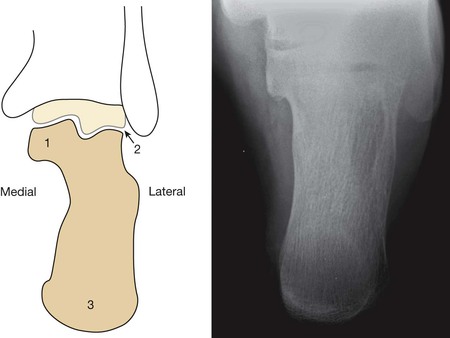
How do you fix a broken calcaneus. A shows the axis of the tibiab shows the surface of the distal tibia and c shows the surface of the proximal talusd shows the line from the top of the sustentaculum tali to the lateral inferior end of the posterior facet of the calcaneuse shows the horizontal line through the. Lateral heel view identifies posterior facet involvement bohlers angle critical angle of gissane and tuberosity position cranial migration.
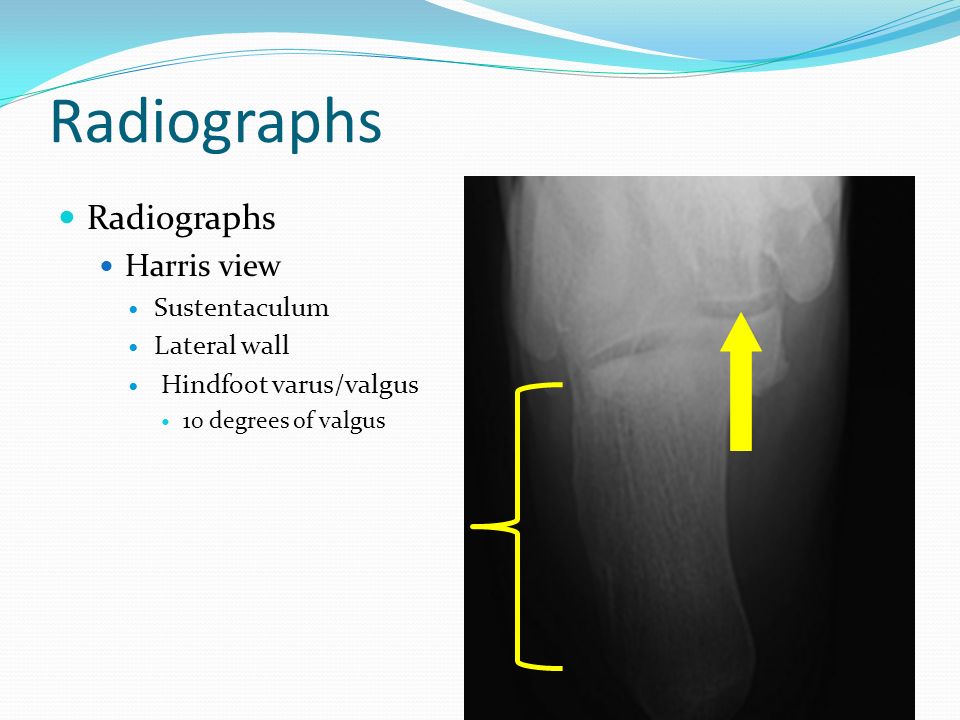
On exam the location of maximal tenderness is indicated by the white arrow in figure a. Note the fracture line divides the calcaneus into anteromedial and posterolateral fragments. Axial view harris beath visualizes tuberosity position varus valgus primary fracture line calcaneal height loss calcaneal widening and a portion of the posterior facet.
As technology advances computed tomography ct has widely been used 1 to better visualize and characterize calcaneum fragment displacements and fracture lines. The ankle joint and the middle and posterior subtalar facets are visualised clearly. The harris view was first described in 1948 by harris and beath as a method of assessing for the presence of a talocalcaneal bridge in a rigid flat foot deformity.
A 48 year old member asked. Sbq12fa56 a 25 year old woman began training for a marathon and she reports a 2 week history of heel pain. The patient stands on the cassette and the x ray beam is angled between 35 and 45 degrees.
40 years experience podiatry. The calcaneus axial view is part of the two view calcaneus series assessing the talocalcaneal joint and plantar aspects of the calcaneus. Yet there remain many institutions especially in rural areas where ct is not readily available.
28 harris axial view of heel. This view is needed for diagnosing the problems of sesamoids figs. The talocalcaneal joint is exposed with the posterior facet laterally and the sustentacular facet medially.
More From Douglas Emhoff Ex Wife Kirsten
- Christmas Donations 2020
- How Old Are Kamala Harris Step Children
- Vp Harris
- Harris Bank Telephone Number
- Biden Campaign Site
Incoming Search Terms:
- Calcaneal Fractures Biden Campaign Site,
- Lower Extremity Plain Radiography Chapter 2 Clinical Emergency Radiology Biden Campaign Site,
- Calcaneum Fracture Pathoanatomy Various Fracture Pattern Biden Campaign Site,
- Calcaneal Fractures Musculoskeletal Key Biden Campaign Site,
- Calcaneus Series Radiology Reference Article Radiopaedia Org Biden Campaign Site,
- Imaging Of The Foot And Ankle Musculoskeletal Key Biden Campaign Site,


