
Harris View X Ray, Wheeless Textbook Of Orthopaedics
Harris view x ray Indeed lately has been hunted by consumers around us, perhaps one of you. People now are accustomed to using the internet in gadgets to view video and image data for inspiration, and according to the name of the article I will discuss about Harris View X Ray.
- Salter Harris Type Iv Fracture Image Radiopaedia Org
- Https Encrypted Tbn0 Gstatic Com Images Q Tbn 3aand9gcrxg800orhb8clhh9lkylpkgvkravthcth5vmqf1jzdxuvxd 8b Usqp Cau
- Imaging Of The Foot And Ankle Musculoskeletal Key
- Paediatric Salter Harris Type Iv Injury Of Distal Tibia With Talus Fracture Bmj Case Reports
- Radiographic Evaluation Harris Heel View Left Is Normal With Parallel Download Scientific Diagram
- Foot Xray Eorif
Find, Read, And Discover Harris View X Ray, Such Us:
- Calcaneus Fracture Extended Lateral Approach Springerlink
- Imaging Of The Foot And Ankle Musculoskeletal Key
- View Image
- Value Of Modified Axial Review Radiograph In Diagnosing Calcaneal Fractures Scientific Reports
- The Acute Occult Salter Harris Injuries In Children
If you are looking for Rocky Kanaka Married you've come to the right place. We ve got 104 graphics about rocky kanaka married including images, pictures, photos, wallpapers, and much more. In such webpage, we also have number of images out there. Such as png, jpg, animated gifs, pic art, symbol, black and white, translucent, etc.

Salter Harris Type Iv Fracture Image Radiopaedia Org Rocky Kanaka Married

Wrist Ap And Lateral View Showing A Salter Harris Fracture Of The Download Scientific Diagram Rocky Kanaka Married

Post Operative Sagittal View A And Harris View B Showing The Download Scientific Diagram Rocky Kanaka Married
Imaging Of The Foot And Ankle Musculoskeletal Key Rocky Kanaka Married
Radiology In Foot And Ankle Musculoskeletal Key Rocky Kanaka Married
Radiology In Foot And Ankle Musculoskeletal Key Rocky Kanaka Married
Pre vertebral soft tissue swelling is also described.
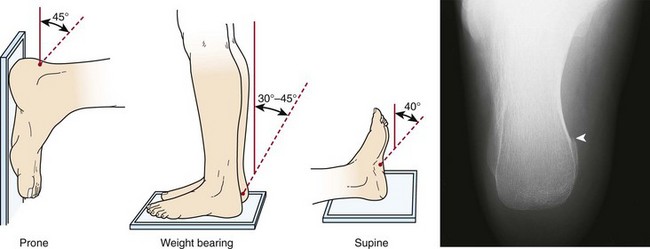
Rocky kanaka married. The talocalcaneal joint is exposed with the posterior facet laterally and the sustentacular facet medially. Harris and beath view. 5 an axial view of the calcaneus is obtained with the x ray source posterior to the heel and tilted caudally 450 with respect to the long axis of the foot.
Fractures of the 5 th metatarsal may also be seen and the medial clear space might be assessed in this view 3. As applied to calcaneal. Fractures of c1 atlas c2 axis c spine vertebral bodies and fracture dislocations are discussed.
We studied 46 feet of individuals with normal appearing asymptomatic feet. Excellent view of posterior subtalar joint given by these views helps in intraoperative monitoring of calcaneus fracture reduction and assessment of. These views are taken with 10 20 30 and 40 degrees cranial angulation of an x ray beam focused at the tip of the fibula with ankle rotated 45 degrees internally.
Trauma patients may not have the ability to rotate their lower limb internally in this case the x ray beam can be angled 15 200 medially to achieve the view although this will result in some artifactual elongation of structures. Harris lines are often discussed as a result. The harris view was first described in 1948 by harris and beath as a method of assessing for the presence of a talocalcaneal bridge in a rigid flat foot deformity.
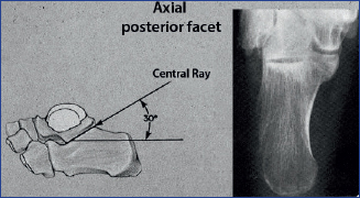
On the antero posterior roentgenogram using this new method the line from the top of the sustentaculum tali to the lateral inferior end of the posterior articular surface of the talus was. Typical fracture patterns include jefferson fracture hangman fracture extension teardrop flexion teardrop perched facet joints and clay shovelers fracture. The x ray beam is aimed from a position slightly distal to the foot with an angle of incidence to the plantar surface of the foot of approximately 30 degrees.
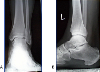
The harris view is obtained with the ankle in dorsiflexion and a facet posteriorly along the course of the achilles tendon. Growth arrest lines also known as harris lines are lines of increased bone density that represent the position of the growth plate at the time of insult to the organism and formed on long bones due to growth arrest. They are only visible by radiograph or in cross section.
The harris projection also called the penetrated axial projection is a special radiographic view that is used for assessment of talocalcaneal coalition. The age at which the lines were formed can be estimated from a radiograph. The patient stands on the cassette and the x ray beam is angled between 35 and 45 degrees.

Ankle X Rays Rocky Kanaka Married

The Acute Occult Salter Harris Injuries In Children Rocky Kanaka Married
Epos Trade Rocky Kanaka Married
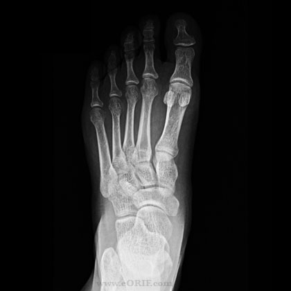
Foot Xray Eorif Rocky Kanaka Married
More From Rocky Kanaka Married
Incoming Search Terms:
- Ce4rt Radiographic Positioning Of The Heel And Ankle For X Ray Techs Vice Comm,
- Ankle Hindfoot Radiology Key Vice Comm,
- Calcaneus Series Radiology Reference Article Radiopaedia Org Vice Comm,
- Radiology In Ped Emerg Med Vol 3 Case 5 Vice Comm,
- Radiology In Ped Emerg Med Vol 4 Case 14 Vice Comm,
- Https Www S3 Live Kent Edu S3fs Root S3fs Public Hv Ch 04 Review Of Adult Foot Radiology Pdf Vice Comm,